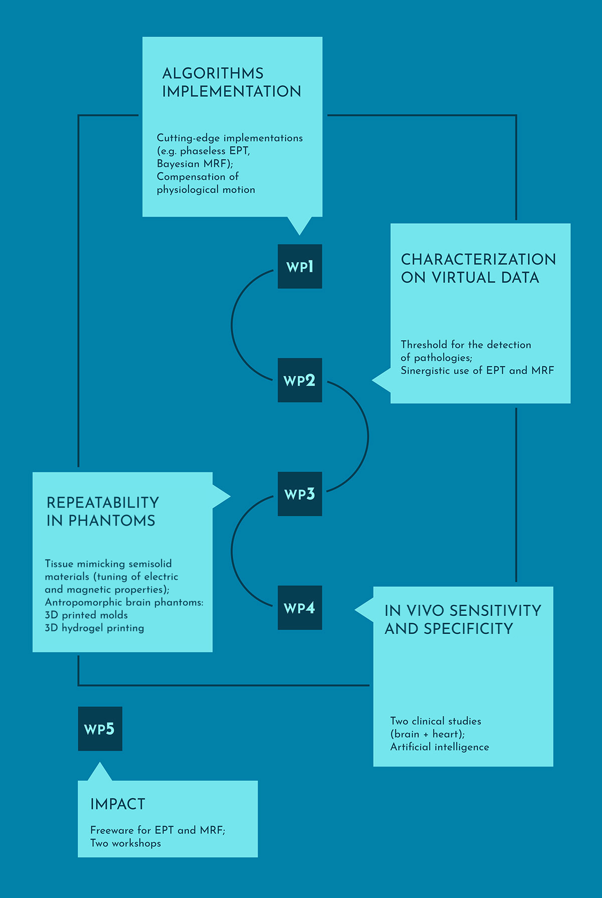
Work package 1
Development of the reconstruction techniques
The aim of this work package is to provide a set of implemented algorithms for EPT and MRF, selected from the most recent literature and developed to include some innovative solutions, capable of overcoming challenging issues (e.g. biased measurements of the transmit sensitivity phase for EPT; physiological motion in cardiac imaging). The WP will be developed with focus on brain and heart imaging, affecting the selection of the B1-mapping sequences and the application of the selected approaches. The techniques will be developed synergistically, exploring the possible employment of MRF approaches to provide fast and accurate inputs to EPT algorithms.
Work package 2
Metrological characterisation of reconstruction techniques in silico
The aim of this work package is to metrologically characterise the quantitative imaging methods for EPT and MRF developed and implemented in WP1, in a completely controlled environment constituted by virtual experiments conducted on realistic in silico models. Moreover, the use of the imaged quantities as physical biomarkers for the presence and/or stage of both localised and diffused pathologies will be investigated.
Virtual experiments will be carried out by solving Maxwell’s and Bloch’s equations sequentially, providing realistic data in completely controlled conditions for the metrological characterisation of EPT and MRF procedures, which, in addition, will allow verifying the feasibility of an MRF-based B1-mapping to be used as an input for EPT. The characterisation will be extended to the detection of anomalies in altered virtual human models imaged with EPT and MRF. Besides traditional statistical approaches, state of the art machine learning techniques will be adopted and tested.
Work package 3
Experimental characterisation of EPT and MRF in phantoms
The target of WP3 is to evaluate the repeatability and reproducibility of the EPT/MRF results under controlled conditions, through experiments involving preclinical and clinical MRI scanners and suitable phantoms whose properties have been characterised via independent methods. To test the ability of EPT/MRF to correctly reproduce tissue parameters even for complex anatomical geometries, phantoms resembling the anatomically correct distribution of different tissue types in the human body will be developed, combining standardised preparation methods for tissue-mimicking materials and the most recent developments in soft-matter and bio-printing. The information gathered in WP3 will be exploited in WP4 to disentangle the variability of results due to varying features of MRI acquisitions from the true physiological variability of tissue properties.
Work package 4
In vivo quantitative differentiation of tissue – towards personalized medicine using metrology and artificial intelligence
The aim of this work package is to apply and test the algorithms for EPT and MRF on in vivo data, from realistic clinical scenarios. The work package will encompass two clinical studies: one for brain, mainly conceived for EPT, and the other for heart diseases, with main focus on MRF. In both clinical studies, both EPT and MRF will be applied to selected patient groups and healthy volunteers. This WP will exploit all the knowledge collected throughout the project in an attempt to (semi) automatically detect clinical anomalies using artificial intelligence (AI). One or more machine learning methods will be modified so as to consider the known uncertainties of its input features when learning and/or when applying the learned model. This will be a stepping-stone towards creating pilot versions of clinical decision support systems using the learned AI models for diagnostic help and easier monitoring of the patients.